Respiratory System
Trachea and Larynx
The larynx, or voice box, contains the vocal chords and is located superiorly to the trachea. To find it, open the thoracic cavity and continue to cut up towards the chin. It is the hard structure at the back of the throat that is connected to the trachea (ringed tube).
The tachea is the windpipe that inhaled air passes through to get to the lungs. It is ringed and rigid and is called the windpipe in contrast to the esophagus, which is composed of smooth muscle. It can be found similar to the larynx cutting from the thoracic cavity up towards the chin. It runs from the larynx to the lungs. |
Thyroid Gland
The thyroid gland lies just above the upper end of the trachea. It's easy to cut through this when first locating the trachea, so be careful. Its main function is to produce hormones for regulating metabolism and lowering blood calcium levels.
To Find it, open the thoracic cavity and start cutting up towards the chin. It is the small, diamond shaped gland that lies right on the trachea or just next to it. |
Caroited Arteries and Aorta
The carotid arteries supply blood to the head and neck and are located symmetrically on either side of the trachea. To find them, first open the thoracic cavity and locate the trachea. They are tubes the look similar to the esophagus, but are a little smaller in width.
The aorta is located at the top of the heart and is responsible for pumping oxygenated blood to the rest of the body. To find it, locate the heart and the large hole at the top is the exit of the aorta. |
Bronchi and Lungs
The bronchi are the little tubes at the end of the trachea that attach the trachea to the lungs. To find them, forst cut into the thoracic cavity. Follow the trachea all the way down to where it meets the lungs and there is a tube connecting the trachea to each lung.
The lungs, like in humans, are responsible for the oxygenation of blood and are essential for breathing. They're pretty easy to find. Start by opening the thoracic cavity and go all the way to the bottom of the trachea. There is one on each side and they're relatively large. The right lung is bigger and contains three lobes (superior, medial, inferior). The left lung is smaller to compensate for the space occupied by the heart. |
Parietal and Visceral Pleura
The pleural membrane lines the small pleural cavity. There are two layers the parietal pleura and the visceral pleura. The shiny membranes are easily identifiable as one that lines the thoracic cavity. The visceral membrane touches the lungs while the other, parietal side touches the thoracic wall.
To find the visceral pleura, simply open up the thoracic cavity and it is the shiny membrane on the thoracic wall. To get at the parietal pleura, you need to peel back the whole membrane. Watch the video for this one and it should be much more clear. |
Thymus
The thymus gland is responsible for development of the immune system and is located near the heart.
To find it just open the thoracic cavity and it is the white with a sponge-like consistency located on the left side of the pig. It might actually overlap the heart a little at the top. It is relatively large compared to the other glands. |
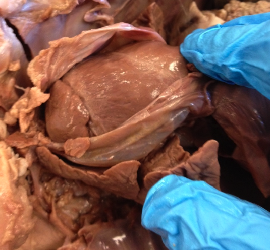
Heart
Pulmonary Trunk, Aorta, Atria, Ventricles, Vena Cava, Pulmonary Arteries, Pulmonary Veins, Coronary VesselesThe pulmonary trunk is an artery that exits the heart on the ventral surface from the right ventricle. It angles to the left and is located on the superior part of the heart, next to the aorta.
The aorta is found behind the large vessle, the pulonary trunk. The part of the aorta leaving the left ventricle is known as the ascending aorta. The aorta appears white and runs parallel with the esophagus, until it reaches the heart. There are two atrias, left and right, on the heart. They cover the top portion of the heart. They are easily seperated from the ventricles because of their dark color. The left atria is actually on the right side when looking at the heart and vice-versa with the right atria. The two ventricles, right and left, are found just bellow the atria. The right and left ventricles are seperated by the coronary vessels that run down the middle of the heart. Like the atria, the right and left ventricles are oppostie when looking at them, right is on the left side and left is on the right side. There are two vena cavas in the heart. The anterior (superior in humans) is a short vessel anterior to the right side of the heart. The posterior (inferior in humans) passes through the diaphram mid-dorsally and is on the ineferior side of the heart. The pulmonary arteries are the parts that are divided from the pulmonary trunk. These can be found by following the pulmonary trunk and finding where it divides into these vessels. The pulmonary veins lie on the superior part where it enters the heart. These are branches of blood vessels that can be traced from the lungs (you will have to lift up the lungs to see them) and enter the heart through the left atria. |
|
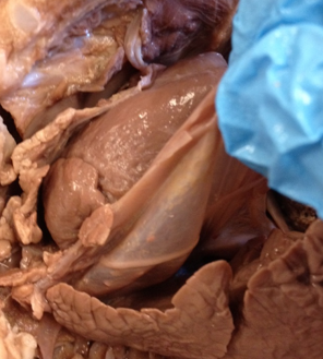
Pericardium
The pericardium, or sac surrounding heart, is a protective membrane enclosing the heart. There is a visceral pericardium that is a thin membrane on the outer surface of the heart and a parietal pericardium that is noticabley thicker.
The pericardium is quite simple to identify. It is a transparent like colored membrane covering the heart. It is possible for the pericardium to be attached to the diaphram as well. |
|
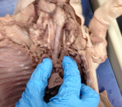
Mediastinum
The Mediastinum is the space located between the lungs. Organs contained within the mediastinum include the heart, esophagus, aorta, and part of the thymus gland. The easiest way to find it is to locate the midline at which the inner thorcic wall forms a partition between the pleural cavities.
The mediastinum is easy to see when the heart is removed and the lungs are pushed off to the side (as shown in picture). |